J.G. Spurzheim, The Anatomy of the Brain, with a General View of the Nervous System. Translated at the request, and under the immediate superintendence of the author from the unpublished French M.S. by R[obert] Willis, MRCS, London, 1826. [Preface & plates only]
 In
this work Spurzheim made bold claims about
what he represented as his own discoveries and techniques in the anatomy of
the brain. How much he in fact borrowed from F.J.
Gall, his long term master, is yet to be demonstrated. Spurzheim also
reproduced in this work portions of text written by Gall in his earlier work.
Below are the 11 plates, many of which Spurzheim took from the 1812 atlas
to Anatomie et physiologie du système nerveux published together
with Gall.
In
this work Spurzheim made bold claims about
what he represented as his own discoveries and techniques in the anatomy of
the brain. How much he in fact borrowed from F.J.
Gall, his long term master, is yet to be demonstrated. Spurzheim also
reproduced in this work portions of text written by Gall in his earlier work.
Below are the 11 plates, many of which Spurzheim took from the 1812 atlas
to Anatomie et physiologie du système nerveux published together
with Gall.
The preface, reproduced below, provides interesting details about Spurzheim's
relations with Gall and claims to independence from the shadow of the more
famous Gall.
Compare Spurzheim's plates with those from William Carpenter's Principles of mental physiology, 1874, Appendix pp. 709-722.
-------------------------
PREFACE.
_____
THE affective and intellectual faculties of man, both in their
healthy and diseased condition, are unquestionably dependent on the body ;
and, among the various branches of anthropology, anatomy is the basis of all
the others. The organic apparatuses, which are indispensable to the mental
manifestations, consist of the brain, cerebellum, spinal cord, and the nerves
of the external senses and voluntary motion. To make known the structure of
these parts is the special object of this volume.
History proves that the structure and destination of the nerves were long
unknown. Hippocrates and Aristotle, for instance, confounded, under the same
general title, ligaments, tendons, nerves, and even blood-vessels. Hippocrates
believed that the nerves terminated in muscles and bones, and produced voluntary
motion. Herophilus, who lived nearly three centuries before the com-
viii PREFACE.
mencement of the vulgar era, was the first who discovered the
connexion of the nerves with the brain, and who looked on them as the instruments
of sensation. Erasistratus divided the nerves into those of sensation and
those of motion; the first he derived from the brain, the second from the
membranes. Galen held that the nerves of sensation arose from the brain, and
those of voluntary motion from the spinal cord. In the sixteenth and succeeding
centuries the brain and nerves were subjects of much research, but it is only
in our own times that they have begun to be understood,-that their true structure
has been discovered, and that new and unthought-of functions have been proved
to belong to them.
The nervous being the most delicate tissues of the body necessarily required
extremely careful and often-repeated examination to be understood, and this
they could not receive during ages when prejudice opposed insurmountable obstacles
to the dissection of dead bodies. It is therefore easy to conceive why such
slow progress was made in the anatomical knowledge of the nervous system.
PREFACE. ix
Dr. Gall is the original author of a new physiological doctrine
of the brain. The discovery of the ground-work of this is all his own, and
he had even gone very far in rearing the superstructure before the year 1804,
when I became his colleague. From this period we continued labouring in common,
until 1813, when our connexion ceased, and each began to pursue the subject
for himself. The works which Dr. Gall has published in his own name fix the
extent of his phrenological knowledge. My ideas, too, are developed in my
own publications: history will assign to each of us his share in the works
that have issued under our joint names.
It was in the year 1800 that I attended for the first time the private course
of lectures which Dr. Gall had been in the habit of delivering occasionally
at his house for four years. At this time he spoke of the necessity of the
brain to the manifestations of mind, of the plurality of the mind's organs,
and of the possibility of discovering the development of the brain by the
configuration of the head. He pointed out several particular organs of different
memories, and of several sentiments, but he had not yet begun to examine the
X PREFACE.
structure of the brain*. Between 1800 and 1804 he modified his
physiological ideas and brought them- to the state in which he professed them
at the commencement of our travels.†
Dr. Gall haying met with a woman, fifty-four years of age, who from her infancy
had laboured under dropsy of the brain, and who, nevertheless, was as active
and intelligent as the generality of females in her own rank of life and being
convinced that the brain was the indispensable organ of the soul, expressed
himself in terms similar to those which Tulpius had used before him, on observing
a person afflicted with hydrocephalus, who exhibited good intellectual faculties,
viz., the structure of the brain must be different from what it is commonly
supposed to be. He now felt the necessity of examining the mind's organ anatomically.
As his medical practice occupied his time, he employed M. Niclas, a student,
to dissect for him ; but the
* Exposition de la Doctrine de Gall, par Froriep, 3me edit,
1802.
† Bischoff Exposition de la Doctrine de M. Gall, sur le Cerceau et le
Crane, Berlin, 1805 ; et Blœde, la Doctrine de Gall Sur les fonctions
du Cerveau, Dresden, 1805,
PREFACE. XI
spirit of this gentleman's researches was merely mechanical,
as is allowed in our joint work, entitled " Anatomie et Physiologie du
Systeme nerveux en général, et du Cerveau en particulier*."
Having completed my studies in 1804, I was associated with Dr. Gall, and devoted
myself especially to anatomical inquiries. At this period, Dr. Gall, in the
Anatomy, spoke of the decussation of the pyramidal bodies, of their passage
through the pons Varolii, of eleven layers of longitudinal and transverse
fibres in the pons, of the continuation of the optic nerve to the anterior
pair of the quadrigeminal bodies, of the exterior bundles of the crura of
the brain diverging beneath the optic nerves in the direction which Vieussens,
Monro, Vicq d'Azyr, and Reil† had followed, the first, by means of scraping,
the others, by cutting the substance of the brain. Dr. Gall shewed further,
the continuation of the anterior commissure across the striated bodies ; he
also spoke of the unfolding of the brain that happens in hydrocephalus. The
notion he had conceived of this, however,
* Preface to the first vol. p 16.
† Gren's Journal, 1795, i. p. 102.
b 2
xii PREFACE.
was not correct ; for he thought that the convolutions resulted
from the duplicature of a membrane, believing that the cerebral crura entered
the hemispheres on one side, expanded there, and then folded back on themselves
by the juxtaposition of the convolutions. The true structure of the convolutions,
and their connexion with the rest of the cerebral mass, were not described
until our joint Memoir was presented to the French Institute in 1808.
The mechanical direction which the anatomical investigations had taken did
not appear to me satisfactory. Guiding myself in my inquiries by physiological
views, always comparing structure with function, I discovered the law of the
successive additions to the cerebral parts ; the divergence in every direction
of the crural bundles towards the convolutions ; the difference between the
diverging fibres and those of union ; the generality of commissures ; the
true connexion of the convolutions with the rest of the cerebral mass, and
the peculiar structure which admits of the convolutions being unfolded (an
event that occurs in hydrocephalus of the cavities), whilst the mass lying
at their bot-
PREFACE. xiii
toms, and belonging, for the most part, to the apparatus of
union, or of the commissures, is pushed by the water between the two layers
composing them ; lastly, I demonstrated the structure of the nervous mass
of the spine, and I flatter myself with having arrived at the best method
of dissecting the brain, and exposing its parts.
What is my object then in publishing this volume ? Our large work is too expensive
for the generality of medical students, and, further, the method pursued in
the discussions there is only calculated for professed anatomists; whilst
this book will be both less costly, and it will be adapted to the student
as well as to the more advanced anatomist. Moreover, many new ideas, possessing
a great share of interest, may now be added; for since Dr. Gall and I first
published, the study of the nervous system has engaged the especial attention
of anatomists and physiologists. I have, myself, continued inquiring, and
conceive that I have made several new discoveries. I have, however, copied
some passages from the first volume of the large work already mentioned, and
also given reduced drawings of several of its
xiv PREFACE.
plates because I think I have acquired a right to this volume,
by its publication in our joint names, by my discoveries that form its principal
object, and by all I did in furtherance of its publication. All the drawings
were executed under my superintendence from anatomical preparations, made
and determined on by me ; the engraver worked by my directions ; no plate
was sent to press without my approval; the descriptions of the plates, and
the anatomical details are mine ; and I furnished the literary notices in
regard to the nerves of the abdominal thorax, to those of the cerebral column,
of the five senses, of the cerebellum, and of the brain.
Whoever desires more copious historical details than this volume will be found
to contain, I refer to our Memoir, addressed to the French Institute, and
to the first volume of our great work, commenced in common, and continued
by Dr. Gall singly, after the middle of the second volume.
The influence our labours have had on the study of the nervous system is incontestable.
To be convinced of this, it is enough to examine the state of knowledge in
regard to the anatomy, physiology, and pathology of the
PREFACE. xv
brain and spinal marrow, when Dr. Gall and I developed our ideas on these matters, whether it was by teaching orally, by dissecting publicly, or by means of our writings. I confess there is great satisfaction in the consciousness of having contributed to the important reform that has been effected in regard to the nervous system. I am only sorry to observe, that many of our ideas are appropriated by the authors of recent publications, without any mention of the source whence they were derived, or of the individuals who first struck them out, or reduced them to certainty by direct proofs. We are commonly enough mentioned, it is true, when such of our assertions as appear weak are the subjects of criticism, but our names are kept in the background when points of importance become the matter of discussion. The public, for instance, by referring to the proper place, may judge whether Mr. J. Cloquet, in his "Anatomy of Man," has been sufficiently explicit in stating, that he has copied every one of the plates of the human brain contained in our large work. M. Serres, whose Memoir was deemed worthy of its prize by the Academy of Sciences of
PREFACE. xvi
Paris, in the first volume of his work, uses our names no fewer
than fifteen times, in connexion with a single idea, which he fancies he can
refute; and generally along with every fact that looks unfavourable to our
opinion, he names us, but he always forgets to cite us in relation to very
many fundamental conceptions which we had announced at the same time. They
who have written to the following effect:—" M. Serres has proved
clearly the erroneousness of M. Gall's observations, and replaced them by
others," may undeceive themselves by attending to the remark I have just
made.
M. Serres's publication forces me likewise to request the reader to distinguish
between a multitude of words and facts on the one hand and the corollaries
which result on the other. I agree with those who, in works of science, pay
especial regard to truths demonstrable to others, to ideas available in practical
life, and to clearness and simplicity of style. To what purpose may serve
the following passage, which occurs in the preliminary discourse of M. Serres,
where, after having said that a monster may be a vegetation of its like, that
it may have,
PREFACE. xvii
two heads, two tails, and six or eight extremities, but that
it would remain strictly confined to the limits of its class, he exclaims
:—" This wonderful phenomenon is undoubtedly connected with the
general harmony of creation. What may be its cause ? We know it not, and in
all likelihood we shall remain ignorant of it for ever. It is one of the mysteries
of creation, whose surface is meted by man, but whose depths are sounded,
and known to God alone *."
This phenomenon does not appear to me more extraordinary than that a kitten
is not a puppy, or that the crab-tree does not produce pears. If the egg of
a bird in its ordinary state cannot produce a mammiferous animal, why should
the germ of this same egg, if it chance to be imperfectly developed, produce
a deformity like to one of the mammalia ? Were the case thus, there would
be some cause for an amazement, but the universal fact of every animal producing
its kind,
* " Cet étonnant phénomène est sans doute hé à l'harmcmîe générale de la création. Quelle peut en être la cause ? Nous l'ignorons et vraisemblablement nous l'ignorerous toujours. C'est un des mystères de la création, dont l'homme mesure la surface, mais dont Dieu &eul sonde et counatt la profondeur."
xviii PREFACE.
is not, in my eyes, more astonishing than any other natural
event.
Further, the mass of facts cited, the number of dissections made, ought never
to impose on us, nor be made a means of concealing the truth. Many of the
anatomists who had lived before us dissected some hundreds of brains, and
they made a boast of their doings in this way ; but they did not perceive
that which I pledge myself to have discovered before I had dissected a dozen
; for instance, the successive additions to the cerebral parts, and the two
kinds of fibres, to wit, the diverging, and the fibres of union. Anatomists
and physiologists had certainly looked upon heads without number ; but before
Dr. Gall's appearance, had failed to discover the seat of a single cerebral
organ. A solitary individual, a beggar, enabled him to detect the organ of
self-esteem, precisely as the fall of a single apple revealed the law of gravitation
to Newton. Anatomists had seen many human brains without remarking any differences
among them; these, however, are, to say the least, as constant as similarities.
The point that essentially interests science is, the
PREFACE. xix
discovery of the truth, and this is then confirmed and established
by all ulterior observations.
The anatomy of the peculiar system necessary to the affective and intellectual
manifestations, as well as anatomy in general, admits of consideration in
several ways.
First, it is simply descriptive, that is, physical appearances alone are examined,
such as the form, the size, and colour of parts, the tissues which compose
them, and their connexions.
The nomenclature of the encephalon, of itself suffices to shew that such views
had principally guided anatomists in their study of its structure. We still
speak of the brain, of the cerebellum, of hemispheres, lobes, convolutions,
and anfractuosities ; of a fornix, an infundibulum or funnel, and of pisiform
and striated, and quadrigeminal and pyramidal, and olivary and harrow-shaped
bodies ; of a pineal gland, of a hippocampus's foot, of a writing-pen, and
many other parts, some with very offensive names. Such views are easily conceived
to be but little useful in medicine. This is the reason why the gene-
xx PREFACE.
ality of practitioners are satisfied with knowing the membraneous
envelopes of the encephalon the large blood-vessels, the sinuses, the great
masses of the brain and cerebellum, and the principal cavities. Of these views,
however, I shall only take such notice as may be necessary to recognize the
parts spoken of in my physiological and pathological considerations.
M. Serres, in the first page of his work on the Anatomy of the Brain, says,
"Up to the present time, (1824,) no one has dreamt of uniting into a
body of doctrine all the knowledge acquired on the anatomy, the physiology
and the pathology of the brain and nervous system. I enter on the attempt
to overtake this vast subject *." Dr. Gall's and my own works belie this
assertion, and they have only to be consulted to prove that all our inquiries
were directed into this very channel. We have constantly insisted on the importance
of studying the nervous system under all relations at once. From the year
1817 to 1823,
* "Jusqu'à ce jour (1824) personne n'a songé à rCunir en corps de doctrine les connaissances acquises sui Panatomie, la physiologie et la pathologie du system nerveux. Je vais essaye de parcourir ce vaste sujet."
PREFACE. xxi
I regularly delivered " a Course of Lectures on the Anatomy,
Physiology, and Pathology of the Brain and External Senses," twice a
year. My course was always so announced, according to the custom in Paris,
by public placards ; and my auditors must recollect that in my introductory
discourse, I uniformly insisted on the importance and necessity of studying
these branches in connexion. From the above, it will be evident that M. Serres
was mistaken when he published himself " the first to attempt to overtake
(essayer de parcourir) this vast subject." Nevertheless, I most willingly
allow that the principal consideration is not the having been the first to
examine the nervous system : the true merit of the inquirer consists in that
which he has effected, that which he had discovered, and justice in these
particulars will, in time, be assuredly tendered to all. If, on occasion,
I seem more especially solicitous in shewing the erroneousness of M. Series's
opinions, it is only because these have received the sanction of the French
Institute, whose influence is great over the public mind.
2. Anatomy is physiological, when the
xxii PREFACE.
structure of parts is studied in relation to their functions.
This kind of anatomical knowledge is essential to practical medicine ; for
without it, the seat of deranged functions cannot be understood. For this
reason, therefore, my anatomical details will always be given in harmony with
the physiological ideas I entertain of the apparatus destined to the manifestation
of the affective and intellectual faculties.
3. Anatomy is peculiarly human, or, it comprehends the other beings of creation.
In the latter event, it is entitled comparative anatomy, and this is a field
that possesses much interest for the anatomist, physiologist, ana practical
physician; I shall, therefore, enter upon it at frequent intervals, always
with the view of advancing the knowledge on the affective and intellectual
nature of man.
4. Anatomy is entitled pathological, when it treats of the organic changes
undergone by parts whether examined in connexion with, or independently of,
their deranged functions. Inquiries in this direction belong less to the present
volume, than to that I have published
PREFACE. xxiii
on Insanity. To it, therefore, I refer the reader for details.
The object of this compendium, then, is to present the principal views that
may be taken of the physiological and comparative anatomy of the apparatus
destined to the affective and intellectual manifestations. It will be found
divided into nine sections:—in the first, I make some general reflections
on the nervous system ; in the second, I speak of the division of the nervous
apparatuses ; in the third, I treat of the nerves of voluntary motion and
of the external senses; in the fourth, I discuss the best mode of examining
the structure of the brain ; in the fifth, I describe the cerebellum particularly
; in the sixth, I do the like in regard to the brain; in the seventh, I examine
the commissures ; in the eighth, the communication of the nervous parts with
each other ; and in the ninth, I go into some anatomical points connected
with physiology. The work will prove that I rather adhere to philosophical
views and principles, than to mere description of the physical qualities belonging
to individual masses, although this last be the most common, I might almost
say
xxiv PREFACE.
the only plan that is generally pursued. I have endeavoured, in an especial manner, to class together the parts that constitute particular apparatuses, a practice which to our predecessors was entirely unknown, as is abundantly evident from their nomenclature of the brain and its parts.
--------------------------
PLATES:
PLATE I.
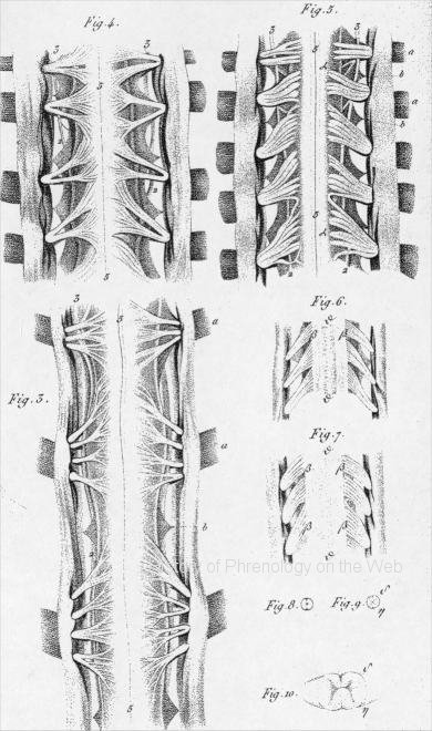
Fig. I. Four ganglions, with their branches of communication, of the caudal
extremity of a caterpillar. This arrangement of the nervous system reigns
through, the entire length, of the body of caterpillars and worms.
Fig. 2. The caudal extremity of the spinal marrow of a fowl, with, the oiigin
of the three last pairs of doi'sal nerves.
Fig. 3. The three superior cervical pairs of nerves in a calf, seen on the
abdominal surface. The dura mater and arachnoid are slit longitudinally
and turned aside, so as to expose the mode in which the nervous filaments
come from the common mass, their different directions, and the passage of
the bundles through the duia mater. The commencement of the intervertébral
ganglions (a) is covered by the reflected membranes. Between the different
pairs of nerves, the teeth of the ligamentum dentatum (e) are seen on each
side of the nervous mass ; these ligaments separate the abdominal fiom the
dorsal roots, and are attached to the dura mater by means of slips, naturally
different in size and position. At the origin of each pair of nerves, a
swelling is perceivable, varying in magnitude, ia proportion to the volume
of the issuing nerve.
PLATE II. -Brains of Fishes
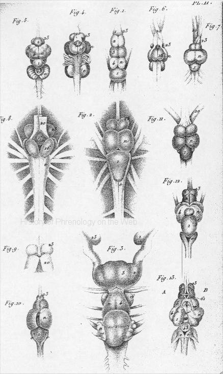
Fig. 1. The brain of an eel seen from above.
Fig. 2. The brain of a cod seen from above.
Fig. 3. The brain of a skate seen from above.
Fig, 4 The brain of a carp seen, from below.
Fig. 5, The brain of a carp seen from above.
Fig. 6. The brain of a Sounder seen from below.
Fig, 7, The brain of a Sounder seen from above,
Fig. 8. The brain of a cod seen from below.
Fig 9. The optic nerves in the cod are thrown back, to permit a view of
the two roots of the olfactory nerves.
Fig. 10, The brain of a pike, -with the cavity of the optic ganglion exposed.
Fig. 11. The brain of a roach.
Fig, 12. The brain of a barbel seen from above.
Fig, 13. The interior of the brain of barbel, prepared to permit a view
of the ganglions, their cotajnunications, and junctions.
PLATE III. -Brains of Reptiles and of Birds.
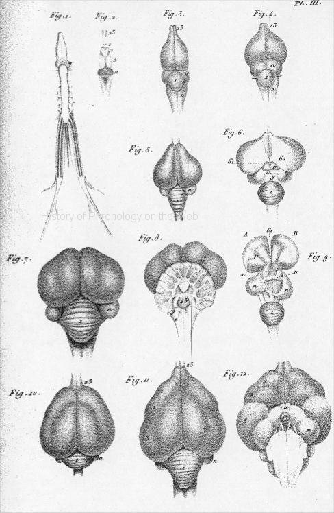
Fig. 1. The spino-cerebial system of a frog seen from below.
Fig. 2. The brain of a frog seen from above.
Fig, 3. From M, Carus's work.-The brain of a lizard (lacerta. iguana) seen
from above.
Fig.4. From M. Carus's work- the brain of a young crocodile seen from above.
Fig.5. The brain of a common fowl seen from above. .
Fig. 6. The brain of a fowl,- The two hemispheres separated. to shew the cerebellum,
the optic .ganglions, 'their commissure, the commencement of the supposed
optic thalimai and the part analogous to the fornix of the mammalia.
Fig. 7. The brain of a turkey seen from above.
Fig.8. The brain of a turkey with the cerebellum cleft-posteriorly, to shew
its lamellar structure,
Fig. 9. The brain of a common fowl, prepared to shew the cavity of the optic
ganglion, the posterior commissure, - the .beginning and continuation of the
great cerebral ganglions.
Fig.10. The brain of a duck seen fcom above.
Fig. 11. The brain of a goose seen from above.
Fig, 12. The brain of a goose seen from below.
PLATE IV. -Brains of Mammiferous Animals.
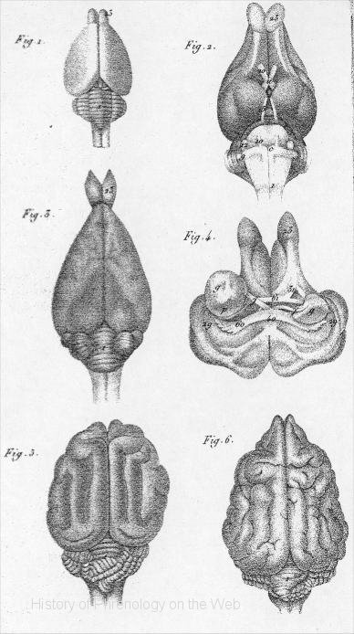
Fig. 1. The brain of myrmesophaga didactyla seen from above,
copied from the work of Tiedemann.
Fig. 2. The brain of a hare seen from below.
Fig, 3. The brain of a hare seen from above.
Fig. 4. The brain of a liare placed on its upper .snriaee, like Fig 2; the
mass below the crura cerebri is cut off ; the crura are separated and pushed
sidewards, to see the under surface of posterior lobes; the fornix, 60; the
posterior fold of the corpus callosum, 40: .the anterior pillar of the fornix
y ; the anterior commisure, 61, .and, its two portions.
Fig. 5. The brain of a cat seen from above.
Fig. 6. The brain of a dog seen from above. All are reduced to the fourth
of their natural size.
PLATE V.
[The brains and their parts, represented in this plate, and all preparations of the human brain, in the following plates, are only a fourth of their natural size.]
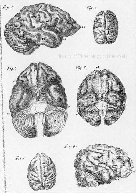
Fig 1. The brain of the monkey, simia saboea, seen from above,
Fig. 2. Tke brain of the monkey, sinnia capucina, seen from above.
Fig. 3. The brain of the ourang-outang seen from below.
Fig. 4. The brain of the ourang-outang seen from the right side.
Fig, 5. The brain of an idiotic girl seen from below.
Fig. 6. The same brain of the idiotic girl seen from the right side.
PLATE VI.
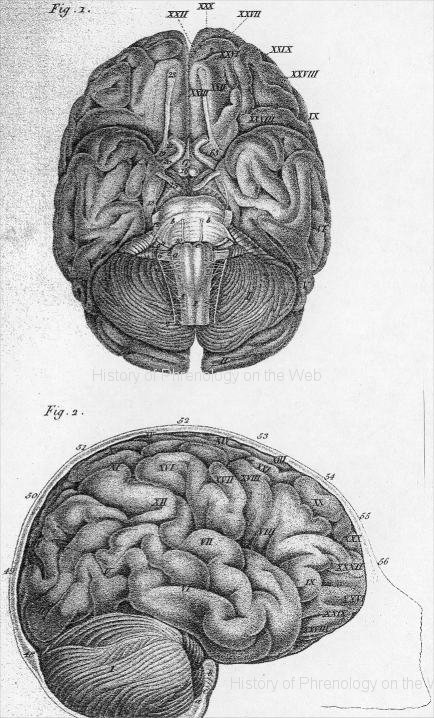
Fig. I. The Basis of the human brain. - All the parts are in
pairs, but not quite symmetrical. The brain is taken from the skull, and turned
on its upper surface; the cerebellum and medulla oblongata have fallen backwards
; the different investing membranes are removed ; the cerebral and nervous
masses alone are visible.
To take-the human brain from the skull, I make an incision from one ear to
the other, turn the integuments backwards and forwards, and detach the temporal
muscles from, the bone. If it is requisite to preserve the skull, it must
be saved about three-quarters of an inch above the superciliary ridge, round
on each, side to the middle of the occipital bone...
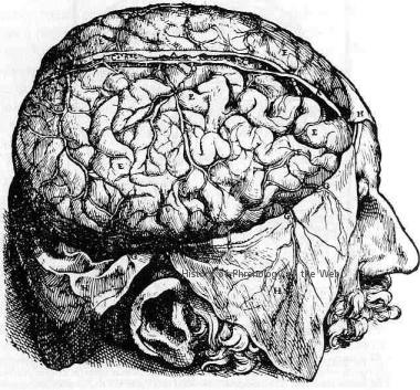
Fig, 2. A skull sawed vertically tbrough the middle of the forehead, the vertex
and the occiput to expose the exterior and lateral surface of the brain, cerebeîlom,
aii-ular protuberance, and medulla oblongata in their Batural situations,
The bone here supports the cerebellum and medulla oblongata, whilst in Fig.
1 they had downwards and backwards.
PLATE VII.
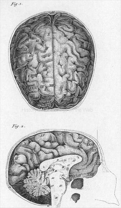
Fig. 1. A skull sawed honsontally in a line from the eye-brows,
by tlie middle oi t fee temples, add the tipper part of the occipital borie.
The membranes and blood-vessels are removed, and the two hemispheres are seen
from above.
Fig. 2, The skull, brain, and cerebellum cut vertically through the median
line and in their natural situations. In this preparation the various parts
are retained in their proper places, not without considerable difficulty.
PLATE VIII.
Fig, 1. A preparation exhibiting various parts about the base
of the brain.
...
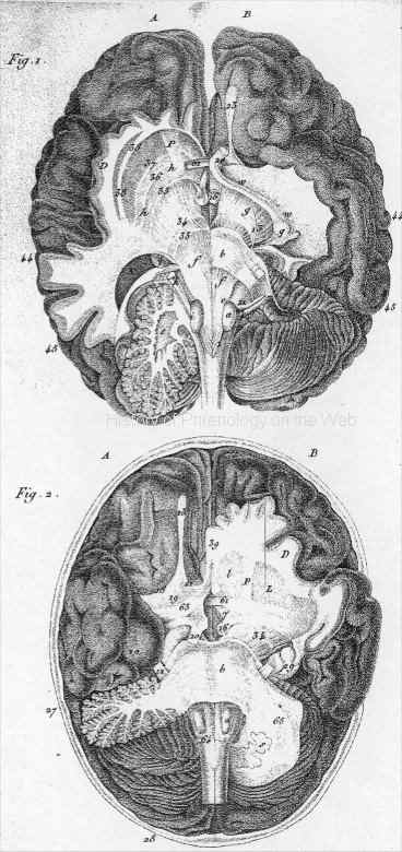
PLATE IX.
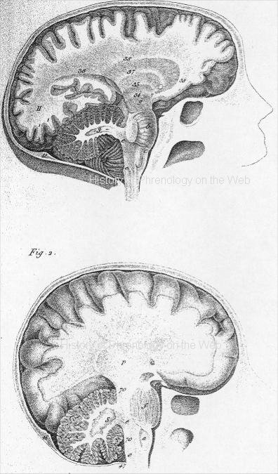
Fig. 1. The cranium sawed on the right side; the cut passes
vertically by the middle of the cerebral and cerebellar hemispheres, and by
the right orbit- The great union of the cerebellum, b, is cut transversely
behind the exit of the trigeminal nerve, and the external part of the cerebellum
is removed. ...
Fig. 2. The cranium sawed perpendicularly in the middle line of the head.
...
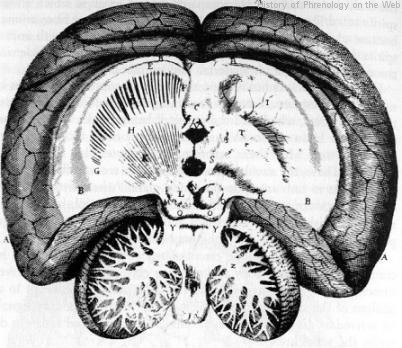
PLATE X.
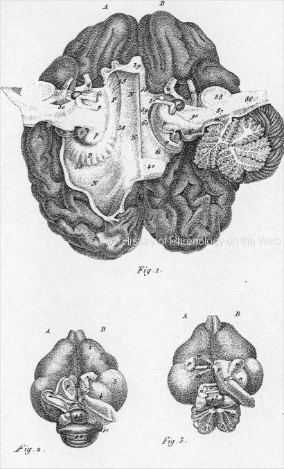
Fig. 1. The brain laid on its superior surface.
PLATE III.
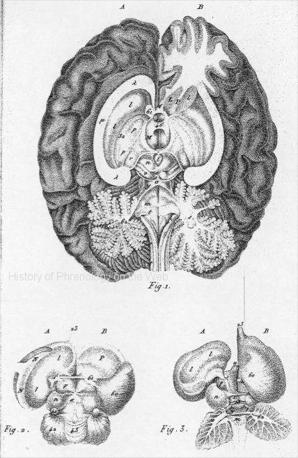
Fig. 1. The human brain laid on its base. ...
----------------------------------------------
See some additional brain images in Combe's System of Phrenology (1853).
Compare Gall and Spurzheim's engravings to those of earlier anatomists.
Vesalius, De Humani Corporus fabrica (1543).
Thomas Willis, De Anima Brutorum (1672).
For more images: Franz Joseph Gall - Johann Gaspar Spurzheim - The Combe brothers
On the brain see especially: GENERAL VIEW OF THE NERVOUS SYSTEM in Combe, System of phrenology, 1850, Carpenter, Principles of mental physiology, 1874, Appendix pp. 709-722.
------------------------
RN3
Digital text and digital images are copyright John van Wyhe 2002.
© John van Wyhe 1999-2011. Materials on this website may not be reproduced without permission except for use in teaching or non-published presentations, papers/theses.